Tag Archives: CD4

First-in-human nanoparticle HIV vaccine induces broad and publicly targeted helper T cell responses

if (g_displayableSlots.mobileTopLeaderboard) {
pushDisplayAd(function() { googletag.display(‘div-gpt-mobile-top-leaderboard’); });
}

Researchers from Fred Hutchinson Cancer Center in Seattle, Scripps Research in La Jolla, California, IAVI and other collaborating institutions have characterized robust T-cell responses in volunteers participating in the IAVI G001 Phase 1 clinical trial to test the safety and immune response of a self-assembling nanoparticle HIV vaccine.

Their work, published in Science Translational Medicine, signals a major step toward development of a vaccine approach to end the HIV/AIDS epidemic worldwide. The antigen used in this study was jointly developed by IAVI and Scripps Research and has been shown in previous analyses to stimulate VRC01-class B cells, an immune response considered promising enough for boosting in further studies.

We were quite impressed that this vaccine candidate produced such a vigorous T-cell response in almost all trial participants who received the vaccine. These results highlight the potential of this HIV-1 nanoparticle vaccine approach to induce the critical T-cell help needed for maturing antibodies toward the pathway of broadly neutralizing against HIV.”

Julie McElrath, MD, PhD, senior vice president and director of Fred Hutch’s Vaccine and Infectious Disease Division and co-senior author of the study

However, she added, this is the first step, and heterologous booster vaccines will still be needed to eventually produce VRC01-class broadly neutralizing antibodies, which in previous studies have demonstrated the ability to neutralize approximately 90% of HIV strains.

“We showed previously that this vaccine induced the desired B-cell responses from HIV broadly neutralizing antibody precursors. Here we demonstrated strong CD4 T-cell responses, and we went beyond what is normally done by drilling down to identify the T cell epitopes and found several broadly immunogenic epitopes that might be useful for developing boosters and for other vaccines,” William Schief, PhD, executive director of vaccine design for IAVI’s Neutralizing Antibody Center at Scripps Research and professor, Department of Immunology and Microbiology, at Scripps Research, who is co-senior author of the study.

if (g_displayableSlots.mobileMiddleMrec) {
pushDisplayAd(function() { googletag.display(‘div-gpt-mobile-middle-mrec’); });
}

The trial is a phase 1, randomized, double-blind and placebo-controlled study to evaluate the safety and effectiveness of a nanoparticle HIV vaccine in healthy adult volunteers without HIV. It was comprised of two groups with 18 vaccine and six placebo recipients per group, with 48 total enrollees. Participants were given two doses of the vaccine or placebo eight weeks apart.

McElrath acknowledged the groundbreaking work of her lab team, the biostatistical team and Fred Hutch’s Vaccine Trials Unit for their invaluable contributions to the study. The Vaccine Trials Unit conducts multiple vaccine trials and was one of only two sites for this study.

Findings from the study include:

  • Vaccine-specific CD4 T cells were induced in almost all vaccine recipients.
  • Lymph node GC T follicular helper cells increased after vaccination compared to placebo.
  • Lumazine synthase protein, needed for self-assembly of the particle, also induced T-cell responses that can provide additional help to ultimately enhance efficacy in a sequential vaccine strategy.
  • Vaccine-specific CD4 T cells were polyfunctional and had diverse phenotypes.
  • LumSyn-specific CD8 T cells were highly polyfunctional and had a predominantly effector memory phenotype.
  • CD4 T-cell responses were driven by immunodominant epitopes with diverse and promiscuous HLA restriction.
  • CD8 T-cell responses to LumSyn were driven by HLA-A*02-restricted immunodominant epitopes B- and T-cell responses correlated within but not between LN and peripheral blood compartments.

This study was funded by the Bill & Melinda Gates Foundation Collaboration for AIDS Vaccine Discovery; IAVI Neutralizing Antibody Center; National Institute of Allergy and Infectious Diseases; and Ragon Institute of MGH, MIT and Harvard.

Study authors WRS and SM are inventors on a patent filed by Scripps and IAVI on the eOD-GT8 monomer and 60-mer immunogens (patent number 11248027, “Engineered outer domain (eOD) of HIV gp 120 and mutants thereof”). WRS, KWC and MJM are inventors on patents filed by Scripps, IAVI and Fred Hutch on immunodominant peptides from LumSyn (Title: Immunogenic compositions; filing no. 63127975).

if (g_displayableSlots.mobileBottomLeaderboard) {
pushDisplayAd(function() { googletag.display(‘div-gpt-mobile-bottom-leaderboard’); });
}

Source:
Journal reference:

Cohen, K. W., et al. (2023) A first-in-human germline-targeting HIV nanoparticle vaccine induced broad and publicly targeted helper T cell responses. Science Translational Medicine. doi.org/10.1126/scitranslmed.adf3309.

Monocytes may be a stable reservoir of HIV in patients taking antiretroviral therapy

To develop treatments that may one day entirely rid the body of HIV infection, scientists have long sought to identify all of the places that the virus can hide its genetic code. Now, in a study using blood samples from men and women with HIV on long-term suppressive therapy, a team led by Johns Hopkins Medicine scientists reports new evidence that one such stable reservoir of HIV genomes can be found in circulating white blood cells called monocytes.

Monocytes are short-lived circulating immune cells that are a precursor to macrophages, immune cells able to engulf and destroy viruses, bacteria and other cells foreign to the host.

In the current research, published March 27 in Nature Microbiology, the scientists found evidence that blood samples from people with HIV undergoing long term, standard antiretroviral therapy contained monocytes that harbor stable HIV DNA capable of infecting neighboring cells.

The scientists say the findings may provide a new direction for efforts to improve therapies and eventually cure HIV, which affects more than 34 million people worldwide, according to the World Health Organization. Current antiretroviral drugs can successfully suppress HIV to nearly undetectable levels, but have not resulted in total eradication of the virus.

We don’t know how critical these monocytes and macrophages are to eradication of HIV, but our results suggest we should continue research efforts to understand their role in this disease.”

Janice Clements, Ph.D., professor of molecular and comparative pathobiology, Johns Hopkins University School of Medicine

Scientists have long known that HIV stashes its genome most often in a type of immune cell called a CD4+ T-cell. These hiding places are known as reservoirs.

“To eradicate HIV, the goal is to find biomarkers for cells that harbor the HIV genome and eliminate those cells,” says Rebecca Veenhuis, Ph.D., assistant professor of molecular and comparative pathobiology at the Johns Hopkins University School of Medicine.

To further study the role of monocytes and macrophages in circulating blood as HIV reservoirs, the Johns Hopkins-led team of scientists obtained blood samples between 2018 and 2022 from 10 men with HIV, all of them taking long-term, standard antiretroviral medications.

The researchers extracted blood cells from the samples and grew the cells in the laboratory. Typically, monocytes transform very quickly -; within about three days -; into macrophages, producing monocyte-derived macrophages.

All 10 men had detectable HIV DNA in their monocytes-turned-macrophages, but at levels 10 times lower than those found in the men’s CD4+ T cells, the well-established HIV reservoir.

For the next phase of the research, to determine if HIV genomes were present in monocytes prior to macrophage differentiation, the team used an experimental assay to detect intact HIV genomes in monocytes. The assay was based on one that fellow Johns Hopkins scientist Robert Siliciano, M.D., Ph.D., developed in 2019 to detect the HIV genome in CD4+ T cells.

The scientists, including research associate Celina Abreu, Ph.D., used the assay on blood samples taken from another group of 30 people (eight men from the first group and 22 female participants) with HIV, also treated with standard antiretroviral therapy. The researchers found HIV DNA in the CD4+ T cells and in monocytes of all 30 participants.

The scientists were also able to isolate HIV produced by infected monocytes from half of the research participants. The virus extracted from these cells was able to infect CD4+ T cells.

Three of the participants had their blood examined several times over the four-year study period, and each time, the scientists found HIV DNA and infectious virus produced by their monocyte-derived macrophages. “These results suggest that monocytes may be a stable reservoir of HIV,” says Clements.

In further research, the Johns Hopkins research team plans to pinpoint the subset of monocytes found to harbor HIV DNA and the source of these infected cells.

Source:
Journal reference:

Veenhuis, R. T., et al. (2023). Monocyte-derived macrophages contain persistent latent HIV reservoirs. Nature Microbiology. doi.org/10.1038/s41564-023-01349-3.

Myeloid cells can harbor HIV in people taking antiretroviral therapy

A subset of white blood cells, known as myeloid cells, can harbor HIV in people who have been virally suppressed for years on antiretroviral therapy, according to findings from a small study supported by the National Institutes of Health. In the study, researchers used a new quantitative method to show that HIV in specific myeloid cells—short-lived monocytes and longer-lived monocyte-derived macrophages—can be reactivated and infect new cells. The findings, published in Nature Microbiology, suggest that myeloid cells contribute to a long-lived HIV reservoir, making these cells an important but overlooked target in efforts to eradicate HIV.

Our findings challenge the prevailing narrative that monocytes are too short-lived to be important in cure efforts. Yes, the cells are short-lived, but our follow-up data show that HIV can persist in monocytes over several years in people who are virally suppressed. The fact that we can detect HIV in these cells over such a long period suggests something is keeping the myeloid reservoir going.”

Rebecca Veenhuis, Ph.D., study author, assistant professor of molecular and comparative pathobiology and of neurology at Johns Hopkins University School of Medicine, Baltimore

The study, led by Veenhuis and colleagues at Johns Hopkins University School of Medicine, was supported by the National Institute of Mental Health, the National Institute of Allergy and Infectious Diseases, and the National Institute on Drug Abuse, all part of NIH.

Antiretroviral medications are effective in treating HIV because they prevent the virus from infecting new cells and multiplying. However, HIV may still exist in cells that are in a resting, or latent, state, creating an HIV reservoir. CD4 T cells, a type of white blood cell, are the most well-studied HIV reservoir. Identifying HIV reservoirs is critical to cure efforts, as latent HIV can be reactivated if people stop taking antiretroviral medications.

Monocytes are immune cells that circulate in the blood for about 3 days before traveling to tissue in various parts of the body, including the brain, where they can mature into macrophages. To date, it has not been clear whether latent HIV in these cells can become active again and infect other cells.

“What’s really important in the long run is understanding how monocytes contribute to the tissue macrophage reservoir,” explained Janice Clements, Ph.D., senior author on the study and professor of molecular and comparative pathobiology at Johns Hopkins University School of Medicine. “If monocytes can carry virus to the brain, or lung, or another part of the body and infect resident macrophages that are self-renewing and live almost indefinitely, that’s a real problem.”  

In the study, Veenhuis, Clements, and colleagues first measured HIV DNA in myeloid cells in a sample of 30 participants with HIV, all of whom were virally suppressed and had been on antiretroviral therapy for at least 5 years. They found detectable levels of HIV genetic material in monocytes and macrophages, though the levels were much lower than those observed in CD4 T cells. In some participants, the HIV genetic material found in monocytes was intact, which suggests it may be capable of infecting other cells if reactivated.

They then used the new quantitative method they developed to directly measure viral spread from HIV found in myeloid cells. The researchers isolated monocytes from blood samples taken from 10 participants and nurtured the monocytes in cultures that contained antiretroviral drugs, to replicate the participants’ baseline physical state. After the monocytes differentiated into macrophages, the researchers introduced an immune activating agent and then added fresh white bloods cells to allow for the virus to spread to new cells.

The researchers collected samples from the cell cultures several times over the next 12 days. They included checkpoints throughout the process to ensure that infected CD4 T cells did not interfere with their measurements.

The results showed that cultures from five of the 10 participants had detectable HIV genetic material in monocyte-derived macrophages that could be reactivated to infect other cells and produce more virus. The participants who had these reactivatable reservoirs of HIV in monocyte-derived macrophages had higher overall levels of HIV DNA material.  

Follow-up data from three participants showed that this reservoir can be long-lived, harboring latent HIV for months to several years. These reservoirs were stable and could be reactivated over time, indicating that monocyte-derived macrophages could contribute to viral rebound if antiretroviral treatment is disrupted.

The researchers note that this study is small and larger studies with more diverse participant pools will be essential to accurately estimate the proportion of people who have latent HIV in myeloid cells. Investigating the mechanisms that replenish the monocyte reservoir over time is a critical next step in this research.

“These findings underscore the importance of broadening the scope of HIV cure efforts,” said Joshua A. Gordon, M.D., Ph.D., director of the National Institute of Mental Health. “Shifting away from a sole focus on CD4 T cells to thinking about CD 4 T cells and myeloid cells together, in context, will propel the field toward more promising strategies for eradicating HIV.”

Source:
Journal reference:

Veenhuis, R. T., et al. (2023). Monocyte-derived macrophages contain persistent latent HIV reservoirs. Nature Microbiology. doi.org/10.1038/s41564-023-01349-3.

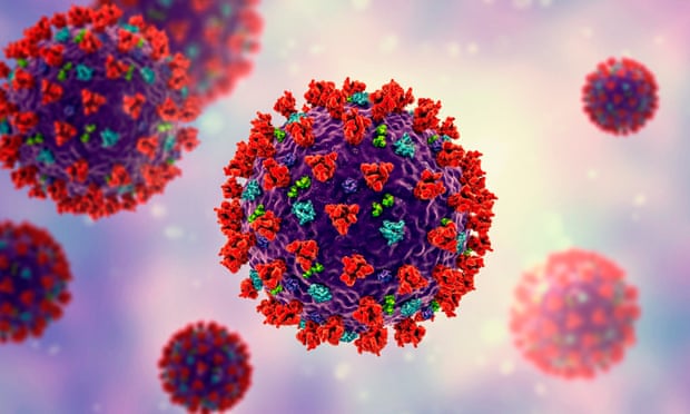
SARS-CoV-2 infection damages the CD8+ T cell response to vaccination

The magnitude and quality of a key immune cell’s response to vaccination with two doses of the Pfizer-BioNTech COVID-19 vaccine were considerably lower in people with prior SARS-CoV-2 infection compared to people without prior infection, a study has found. In addition, the level of this key immune cell that targets the SARS-CoV-2 spike protein was substantially lower in unvaccinated people with COVID-19 than in vaccinated people who had never been infected. Importantly, people who recover from SARS-CoV-2 infection and then get vaccinated are more protected than people who are unvaccinated. These findings, which suggest that the virus damages an important immune-cell response, were published today in the journal Immunity.

The study was co-funded by the National Institute of Allergy and Infectious Diseases (NIAID), part of the National Institutes of Health, and led by Mark M. Davis, Ph.D. Dr. Davis is the director of the Stanford Institute for Immunity, Transplantation and Infection and a professor of microbiology and immunology at Stanford University School of Medicine in Palo Alto, California. He is also a Howard Hughes Medical Institute Investigator.

Dr. Davis and colleagues designed a very sensitive tool to analyze how immune cells called CD4+ T cells and CD8+ T cells respond to SARS-CoV-2 infection and vaccination. These cells coordinate the immune system’s response to the virus and kill other cells that have been infected, helping prevent COVID-19. The tool was designed to identify T cells that target any of dozens of specific regions on the virus’s spike protein as well as some other viral regions. The Pfizer-BioNTech vaccine uses parts of the SARS-CoV-2 spike protein to elicit an immune response without causing infection.

The investigators studied CD4+ and CD8+ T-cell responses in blood samples from three groups of volunteers. One group had never been infected with SARS-CoV-2 and received two doses of the Pfizer-BioNTech COVID-19 vaccine. The second group had previously been infected with SARS-CoV-2 and received two doses of the vaccine. The third group had COVID-19 and was unvaccinated.

The researchers found that vaccination of people who had never been infected with SARS-CoV-2 induced robust CD4+ and CD8+ T-cell responses to the virus’ spike protein. In addition, these T cells produced multiple types of cell-signaling molecules called cytokines, which recruit other immune cells—including antibody-producing B cells—to fight pathogens. However, people who had been infected with SARS-CoV-2 prior to vaccination produced spike-specific CD8+ T cells at considerably lower levels—and with less functionality—than vaccinated people who had never been infected. Moreover, the researchers observed substantially lower levels of spike-specific CD8+ T cells in unvaccinated people with COVID-19 than in vaccinated people who had never been infected.

Taken together, the investigators write, these findings suggest that SARS-CoV-2 infection damages the CD8+ T cell response, an effect akin to that observed in earlier studies showing long-term damage to the immune system after infection with viruses such as hepatitis C or HIV. The new findings highlight the need to develop vaccination strategies to specifically boost antiviral CD8+ T cell responses in people previously infected with SARS-CoV-2, the researchers conclude.  

Source:
Journal reference:

Gao, F., et al. (2023). Robust T cell responses to Pfizer/BioNTech vaccine compared to infection and evidence of attenuated peripheral CD8+ T cell responses due to COVID-19. Immunity. doi.org/10.1016/j.immuni.2023.03.005.

Analysis of rebound virus suggests two separate reservoirs of latent HIV in patients

When people living with HIV take antiviral therapy (ART), their viral loads are driven so low that a standard blood test cannot detect the virus. However, once ART is stopped, detectable HIV re-emerges with new cells getting infected. This is called “rebound” virus, and the cells that release the virus to re-ignite the infection come from a small population of HIV-infected CD4+ T cells that had remained dormant in blood and lymph tissue while individuals were on ART.

It’s a problem called latency, and overcoming it remains a major goal for researchers trying to create curative therapies for HIV-;the special focus of the UNC HIV Cure Center.

Now, scientists led by virologist Ron Swanstrom, PhD, Director of the UNC Center for AIDS Research and the Charles P. Postelle, Jr. Distinguished Professor of Biochemistry & Biophysics at the UNC School of Medicine, describe another layer to the challenge of HIV latency and published their work in Nature Microbiology.

Swanstrom and colleagues, with collaborators at UCSF, Yale, the University of Gothenburg in Sweden, and others, provide indirect evidence for the existence of a distinct latent reservoir of CD4+ T cells in the central nervous system (CNS). They accomplished this by analyzing rebound virus in the cerebral spinal fluid (CSF) during the period when people had just stopped taking ART.

Our analysis of rebound virus suggests latently infected T cells in the CNS are separate from the latent reservoir in the blood. Our analysis allows us to infer the presence of a distinct pool of latently infected cells in the CNS waiting to reinitiate infection once ART is interrupted.”

Dr. Ron Swanstrom, senior author of the study

The researchers compared the genetic sequences of rebound virus particles when ART was discontinued in 11 human participants. This approach allowed the scientists to assess the similarities between viral populations in the blood and CSF to determine whether they were part of a common latent reservoir. In many cases, the viral populations were not the same, which suggested they can represent different populations of latently infected cells.

The researchers also studied details of viral replication to determine if rebound virus had been selected for replication in CD4+ T cells – the primary home of the virus – or had evolved to replicate in central nervous system myeloid cells, such as macrophages and microglia. All rebound viruses tested were adapted to growth in T cells. For several participants, the researchers also compared viral populations in blood and CSF before ART initiation and after ART was stopped.

These experiments provide further evidence that HIV-infected CD4+ T cells can cross over from blood into the CNS, but also that some latently infected cells may be resident in the CNS during therapy. Any curative therapy would need to activate this dormant reservoir, as well as the latent reservoir in the blood and lymph tissue.

Source:
Journal reference:

Swanstrom, R., et al. (2022) Rebound virus in the cerebrospinal fluid reveals a possible HIV-1 reservoir. Nature Microbiology. doi.org/10.1038/s41564-022-01309-3.

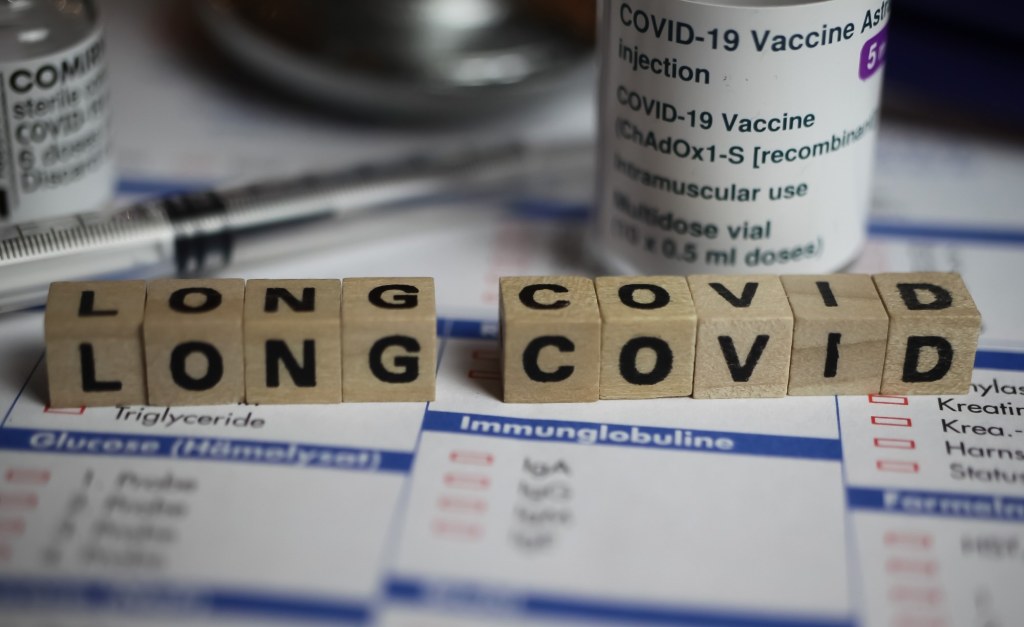
What are the major findings of long COVID research?

In a recent review published in Nature Reviews Microbiology, researchers explored existing literature on long coronavirus disease (COVID). They highlighted key immunological findings, similarities with other diseases, symptoms, associated pathophysiological mechanisms, and diagnostic and therapeutic options, including coronavirus disease 2019 (COVID-19) vaccinations.

Study: Long COVID: major findings, mechanisms and recommendations. Image Credit: Ralf Liebhold/Shutterstock

Long COVID refers to a multisystemic disease among SARS-CoV-2 (severe acute respiratory syndrome coronavirus 2)-positive individuals, with increasing prevalence rates by the day. Studies have reported on long COVID risk factors, symptoms, pathophysiology, diagnosis, and treatment options, with increasing similarities between long COVID and other diseases such as POTS (postural orthostatic tachycardia syndrome) and ME/CFS (myalgic encephalomyelitis/ chronic fatigue syndrome).

About the review

In the present review, researchers explored the existing data on long COVID immunology, symptoms, pathophysiology, diagnosis, and therapeutic options.

Key long COVID findings and similarities with other diseases

Studies have reported persistently reduced exhausted T lymphocytes, dendritic cells, cluster of differentiation 4+ (CD4+) lymphocyte and CD8+ lymphocyte counts, and greater PD1 (programmed cell death protein-1) expression. In addition, increase in innate cell immunological activities, non-classical monocytes, expression of interferons (IFNs)-β, λ1, and interleukins (IL)-1β, 4,6, tumor necrosis factor (TNF). Cytotoxic T lymphocyte expansion has been linked to gastrointestinal long COVID symptoms, and persistent increase in CCL11 (C-X-C motif chemokine 11) expression has been linked to cognitive dysfunction among long COVID patients.

Elevated autoantibody titers have been reported among long COVID patients, such as autoantibodies against ACE2 (angiotensin-converting enzyme 2), angiotensin II receptor type I (AT1) receptors, β2-adrenoceptors, angiotensin 1–7 Mas receptors, and muscarinic M2 receptors. Reactivation of Epstein-Barr virus (EBV) and human herpes virus-6 (HHV-6) has been reported in long COVID patients and ME/CFS. EBV reactivation has been linked to neurocognitive impairments and fatigue in long COVID.

SARS-CoV-2 persistence reportedly drives long COVID symptoms. SARS-CoV-2 proteins and/or ribonucleic acid (RNA) have been detected in cardiovascular, reproductive, cranial, ophthalmic, muscular, lymphoid, hepatic, and pulmonary tissues, and serum, breast, urine, and stool obtained from long COVID patients. Similar immunological patterns are noted between long COVID and ME/CFS, with elevated cytokine levels in the initial two to three years of disease, followed by reduction with time, without symptomatic improvements in ME/CFS. Lower cortisol levels, mitochondrial dysfunction, post-exertional malaise, dysautonomia, mast cell activation, platelet hyperactivation, hypermobility, endometriosis, menstrual alterations, and intestinal dysbiosis occur in both conditions.

Long COVID symptoms and underlying pathophysiological mechanisms

Long COVID-associated organ damage reportedly results from COVID-19-induced inflammation and associated immune responses. Cardiovascular long COVID symptoms such as chest pain and palpitations have been associated with endothelial dysfunction, micro-clotting, and lowered vascular density. Long COVID has been associated with an increased risk of renal damage and type 2 diabetes. Ophthalmic symptoms of long COVID, including altered pupillary responses to light, result from the loss of small nerve fibers in the cornea, increased dendritic cell density, and impaired retinal microvasculature. Respiratory symptoms such as persistent cough and breathlessness result from altered pulmonary perfusion, epithelial injury, and air entrapment in the airways.

Cognitive and neurological long COVID symptoms include loss of memory, cognitive decline, sleep difficulties, paresthesia, balancing difficulties, noise and light sensitivity, tinnitus, and taste and/or smell loss. Underlying pathophysiological mechanisms include kynurenine pathway activation, endothelial injury, coagulopathy, lower cortisol levels, loss of myelin, microglial reactivation, oxidative stress, hypoxia, and tetrahydrobiopterin deficiency.  Gastrointestinal symptoms such as pain in the abdomen, nausea, appetite loss, constipation, and heartburn have been associated with elevated Bacteroides vulgatus and Ruminococcus gnavus counts and lower Faecalibacterium prausnitzii counts. Neurological symptoms often have a delayed onset, worsen with time and persist longer than respiratory and gastrointestinal symptoms, and long COVID presents similarly in children and adults.

Diagnostic and therapeutic options for long COVID, including COVID-19 vaccines

The diagnosis and treatment of long COVID are largely symptom-based, including tilt tests for POTS, magnetic resonance imaging (MRI) to detect cardiovascular and pulmonary impairments, and electrocardiograms to detect QRS complex fragmentation. Salivary tests and serological tests, including red blood cell deformation, lipid profile, complete blood count, D-dimer, and C-reactive protein (CRP) evaluations, can be performed to assess immunological biomarker levels. PCR (polymerase chain reaction) analysis is used for SARS-CoV-2 RNA detection and quantification, and antibody testing is performed to assess humoral immune responses against SARS-CoV-2.

Pharmacological treatments include intravenous Ig for immune dysfunction, low-dosage naltrexone for neuronal inflammation, beta-blockers for POTS, anticoagulants for microclot formation, and stellate ganglion blockade for dysautonomia. Other options include antihistamines, paxlovid, sulodexide, and pycnogenol. Non-pharmacological options include cognitive pacing for cognitive impairments, diet limitations for gastrointestinal symptoms, and increasing salt consumption for POTS. COVID-19 vaccines have conferred minimal protection against long COVID, the development of which depends on the causative SARS-CoV-2 variant, and the number of vaccination doses received. Long COVID has been reported more commonly post-SARS-CoV-2 Omicron BA.2 subvariant infections.

Based on the review findings, long COVID is a multiorgan disease that has debilitated several lives worldwide, for which diagnostic and therapeutic options are inadequate. The findings underscored the need for future studies, clinical trials, improved education, mass communication campaigns, policies, and funding to reduce the future burden of long COVID.

Journal reference: