Microbiom News
News
Microbiology
Microbiome
Health u0026amp; Nutrition
Diseases u0026amp; Disorders
Infectious diseases
Product Review
MARINE MICROBIOLOGY
ANTIMICROBIALS
VIRAL INFECTION
EPIDEMIOLOGY
TOPICS
Microbiome
Health u0026amp; Nutrition
Diseases u0026amp; Disorders
Infectious diseases
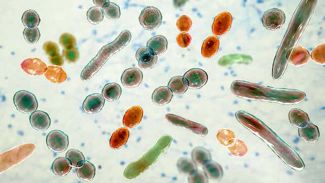
Bacteria
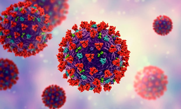
Virus
COVID-19
antibiotic resistance
Gastroenterology
Pathogen
Microbials
Antibiotics
Vaccinations
Microbiome
Health & Nutrition
Diseases & Disorders
Product Review
Newsletter
Contact
Facebook
Instagram
TikTok
Pinterest
YouTube
Twitter
Microbiome & Microbiology Review
Search
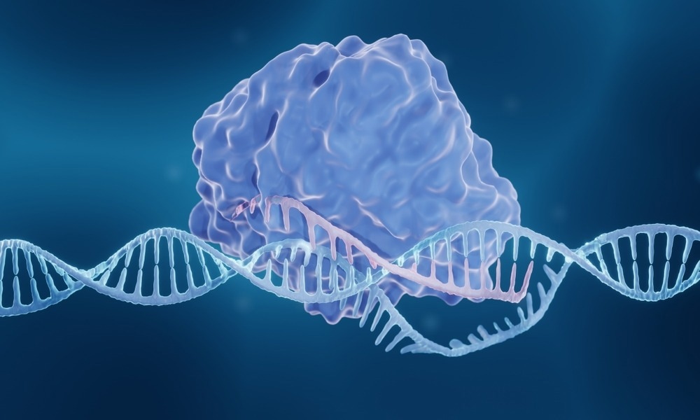
CRISPR
25 May, 2023
New tool shows early promise to help reduce the spread of antimicrobial resistance
19 May, 2023
Novel gene-editing strategy harnesses an unusual protective ability to eliminate HIV-1 infection
13 May, 2023
Study identifies key genetic mechanism of drug resistance in the deadliest malaria parasites
29 April, 2023
Fighting tuberculosis with the new MTB Strip Test Kit
22 March, 2023
“Glow-in-the-Dark” Proteins: The Future of Viral Disease Detection?
21 March, 2023
Avanced genome editing technology could be used as a one-time treatment for CD3 delta SCID
14 January, 2023
Targeting T cell iron metabolism may offer a new approach for treating lupus
11 January, 2023
Newly discovered CRISPR immune system shuts down infected cells to thwart infection
10 January, 2023
Scientists Successfully Edit the Genes of Nature’s Master Manipulators
6 January, 2023
Revolutionary Cancer Vaccine Simultaneously Kills and Prevents Brain Tumors
Next Page
→
Subscribe
Subscribed
Microbiome & Microbiology Review
Join 108 other subscribers
Sign me up
Already have a WordPress.com account?
Log in now.
Microbiome & Microbiology Review
Edit Site
Subscribe
Subscribed
Sign up
Log in
Report this content
View site in Reader
Manage subscriptions
Collapse this bar