Microbiom News
News
Microbiology
Microbiome
Health u0026amp; Nutrition
Diseases u0026amp; Disorders
Infectious diseases
Product Review
MARINE MICROBIOLOGY
ANTIMICROBIALS
VIRAL INFECTION
EPIDEMIOLOGY
TOPICS
Microbiome
Health u0026amp; Nutrition
Diseases u0026amp; Disorders
Infectious diseases
Bacteria
Virus
COVID-19
antibiotic resistance
Gastroenterology
Pathogen
Microbials
Antibiotics
Vaccinations
Microbiome
Health & Nutrition
Diseases & Disorders
Product Review
Newsletter
Contact
Facebook
Instagram
TikTok
Pinterest
YouTube
Twitter
Microbiome & Microbiology Review
Search
Genome
10 June, 2023
Built Into the Genome of the Microbes – Scientists Uncover Over 30,000 “Hidden” Viruses
7 June, 2023
Differences in tumor mutation burden are a major reason for divergence: Study
5 June, 2023
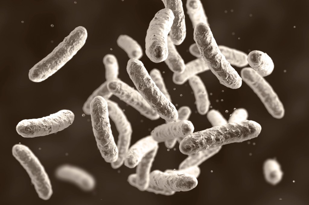
Escherichia coli ST2797 as a novel emerging extended-spectrum beta-lactamase Escherichia coli
2 June, 2023
Experts find remnants of ancient RNA viruses embedded inside reef-building corals
25 May, 2023
Identifying what makes some gut bacteria strains life-threatening in pre-term babies
17 May, 2023
A novel approach to quantify personal information contained within gut metagenome data
13 May, 2023
Study identifies key genetic mechanism of drug resistance in the deadliest malaria parasites
10 May, 2023
MGI Empowers the Completion of Nearly 60,000 Samples for The Million Microbiome of Humans Project
4 May, 2023
Reconstructing ancient bacterial genomes can revive previously unknown molecules – offering a potential source for new antibiotics
4 May, 2023
How the COVID pandemic has improved genomics
Next Page
→
Subscribe
Subscribed
Microbiome & Microbiology Review
Join 108 other subscribers
Sign me up
Already have a WordPress.com account?
Log in now.
Microbiome & Microbiology Review
Edit Site
Subscribe
Subscribed
Sign up
Log in
Report this content
View site in Reader
Manage subscriptions
Collapse this bar