Microbiom News
News
Microbiology
Microbiome
Health u0026amp; Nutrition
Diseases u0026amp; Disorders
Infectious diseases
Product Review
MARINE MICROBIOLOGY
ANTIMICROBIALS
VIRAL INFECTION
EPIDEMIOLOGY
TOPICS
Microbiome
Health u0026amp; Nutrition
Diseases u0026amp; Disorders
Infectious diseases
Bacteria
Virus
COVID-19
antibiotic resistance
Gastroenterology
Pathogen
Microbials
Antibiotics
Vaccinations
Microbiome
Health & Nutrition
Diseases & Disorders
Product Review
Newsletter
Contact
Facebook
Instagram
TikTok
Pinterest
YouTube
Twitter
Microbiome & Microbiology Review
Search
Metabolism
8 June, 2023
Intestinal Bacteria – The Secret to Living to 100?
1 June, 2023
Unique combination of intestinal bacteria in Japanese centenarians may be the key to long life
19 May, 2023
Transforming antibiotic resistance testing: a novel, rapid and affordable technique
27 April, 2023
Discovery of Helicobacter’s Achilles heel offers great potential for the development of new drugs
19 April, 2023
Can a disrupted gut microbiota contribute to anorexia nervosa pathogenesis?
18 April, 2023
Study finds sugary beverages increase dementia risk, while natural juices may help prevent it
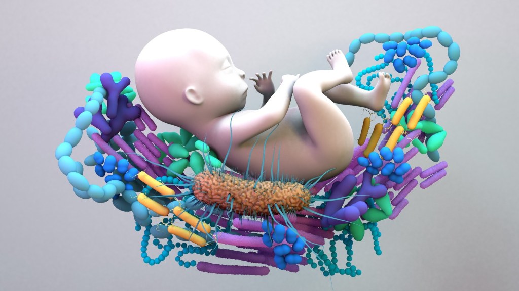
18 April, 2023
Factors shaping maternal gut microbiome during pregnancy and the impact on infant health
4 April, 2023
Live attenuated nasal vaccine elicits superior immunity to SARS-CoV-2 variants in hamsters
29 March, 2023
Study offers a novel therapeutic option to combat antibiotic-resistant pneumonia
23 March, 2023
Novel subset of memory B cells predicts long-lived antibody responses to influenza vaccination
Next Page
→
Subscribe
Subscribed
Microbiome & Microbiology Review
Join 108 other subscribers
Sign me up
Already have a WordPress.com account?
Log in now.
Microbiome & Microbiology Review
Edit Site
Subscribe
Subscribed
Sign up
Log in
Report this content
View site in Reader
Manage subscriptions
Collapse this bar