Microbiom News
News
Microbiology
Microbiome
Health u0026amp; Nutrition
Diseases u0026amp; Disorders
Infectious diseases
Product Review
MARINE MICROBIOLOGY
ANTIMICROBIALS
VIRAL INFECTION
EPIDEMIOLOGY
TOPICS
Microbiome
Health u0026amp; Nutrition
Diseases u0026amp; Disorders
Infectious diseases
Bacteria
Virus
COVID-19
antibiotic resistance
Gastroenterology
Pathogen
Microbials
Antibiotics
Vaccinations
Microbiome
Health & Nutrition
Diseases & Disorders
Product Review
Newsletter
Contact
Facebook
Instagram
TikTok
Pinterest
YouTube
Twitter
Microbiome & Microbiology Review
Search
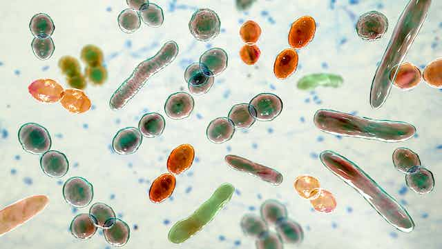
antibiotic resistance
9 June, 2023
Global study provides high-quality data to improve treatment of newborn babies with sepsis
8 June, 2023
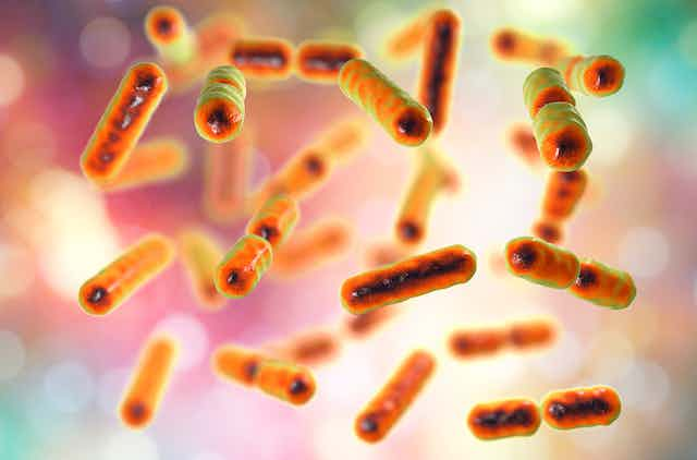
Previously unknown antibiotic resistance widespread among bacteria
7 June, 2023
Global response to antimicrobial resistance ‘insufficient’
7 June, 2023
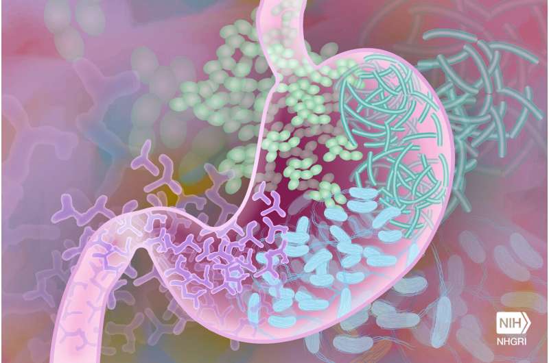
How do antibiotics affect the ecology of the gut microbiome?
6 June, 2023
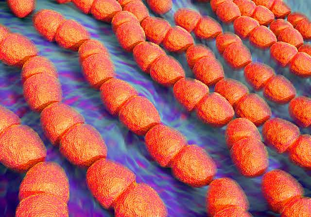
New class of antibiotics to fight resistant bacteria
5 June, 2023
Escherichia coli ST2797 as a novel emerging extended-spectrum beta-lactamase Escherichia coli
5 June, 2023
An overview of the French antibiotic resistance surveillance system in the human, animal, and environmental sectors
25 May, 2023
Failed antibiotic now a game changing weed killer for farmers
22 May, 2023
How superbug A. baumannii survives metal stress and resists antibiotics
19 May, 2023
Transforming antibiotic resistance testing: a novel, rapid and affordable technique
Next Page
→
Subscribe
Subscribed
Microbiome & Microbiology Review
Join 111 other subscribers
Sign me up
Already have a WordPress.com account?
Log in now.
Microbiome & Microbiology Review
Edit Site
Subscribe
Subscribed
Sign up
Log in
Report this content
View site in Reader
Manage subscriptions
Collapse this bar