Microbiom News
News
Microbiology
Microbiome
Health u0026amp; Nutrition
Diseases u0026amp; Disorders
Infectious diseases
Product Review
MARINE MICROBIOLOGY
ANTIMICROBIALS
VIRAL INFECTION
EPIDEMIOLOGY
TOPICS
Microbiome
Health u0026amp; Nutrition
Diseases u0026amp; Disorders
Infectious diseases
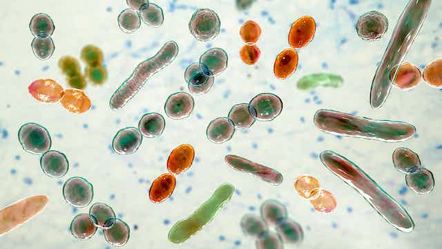
Bacteria
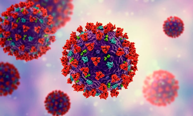
Virus
COVID-19
antibiotic resistance
Gastroenterology
Pathogen
Microbials
Antibiotics
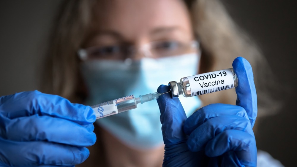
Vaccinations
Microbiome
Health & Nutrition
Diseases & Disorders
Product Review
Newsletter
Contact
Facebook
Instagram
TikTok
Pinterest
YouTube
Twitter
Microbiome & Microbiology Review
Search
Antibodies
31 May, 2023
Experimental decoy provides long-term protection from SARS-Cov-2 infection
25 May, 2023
First-in-human nanoparticle HIV vaccine induces broad and publicly targeted helper T cell responses
19 May, 2023
Mouse study offers clues to developing an effective vaccine for Klebsiella bacteria
17 May, 2023
Novel antibodies target human receptors to neutralize SARS-CoV-2 variants and future sarbecoviruses
8 May, 2023
Anticoronavirals: the development of COVID-19 therapies and the challenges that remain
27 April, 2023
Long COVID: Cedars-Sinai Researchers Find COVID-19 Vaccine Produces Antibodies Far Longer Than Expected
24 April, 2023
Bioengineered drug candidate can counter S. aureus infection in early tests
12 April, 2023
A creative new approach to make vaccine against norovirus
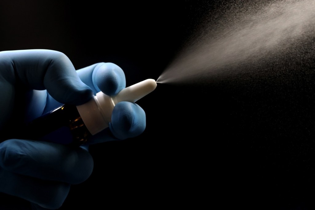
7 April, 2023
Nasal SARS-CoV-2 vaccine outperforms existing vaccines in preclinical trial
6 April, 2023
The Immunity Puzzle: Why Viruses Like COVID-19 Can Reinfect Hosts, Evade the Immune Response
Next Page
→
Subscribe
Subscribed
Microbiome & Microbiology Review
Join 108 other subscribers
Sign me up
Already have a WordPress.com account?
Log in now.
Microbiome & Microbiology Review
Edit Site
Subscribe
Subscribed
Sign up
Log in
Report this content
View site in Reader
Manage subscriptions
Collapse this bar