Microbiom News
News
Microbiology
Microbiome
Health u0026amp; Nutrition
Diseases u0026amp; Disorders
Infectious diseases
Product Review
MARINE MICROBIOLOGY
ANTIMICROBIALS
VIRAL INFECTION
EPIDEMIOLOGY
TOPICS
Microbiome
Health u0026amp; Nutrition
Diseases u0026amp; Disorders
Infectious diseases
Bacteria
Virus
COVID-19
antibiotic resistance
Gastroenterology
Pathogen
Microbials
Antibiotics
Vaccinations
Microbiome
Health & Nutrition
Diseases & Disorders
Product Review
Newsletter
Contact
Facebook
Instagram
TikTok
Pinterest
YouTube
Twitter
Microbiome & Microbiology Review
Search
Bacteria
25 August, 2023
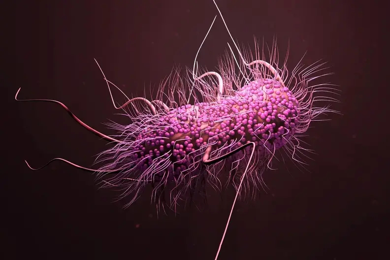
Urgent Steps Required to Combat the Spread of Superbugs and Antimicrobial Resistance, Says UN Report
8 June, 2023
Gut microbiome changes linked to precancerous colon polyps
7 June, 2023
How do antibiotics affect the ecology of the gut microbiome?
7 June, 2023
AI Revolutionizes Antibiotic Discovery: A New Hope Against Evasive Hospital Superbugs
6 June, 2023
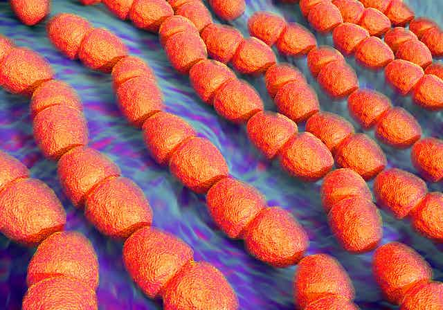
New class of antibiotics to fight resistant bacteria
5 June, 2023
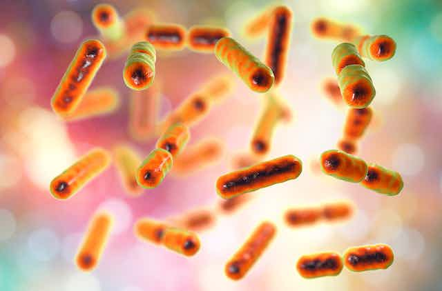
Escherichia coli ST2797 as a novel emerging extended-spectrum beta-lactamase Escherichia coli
4 June, 2023
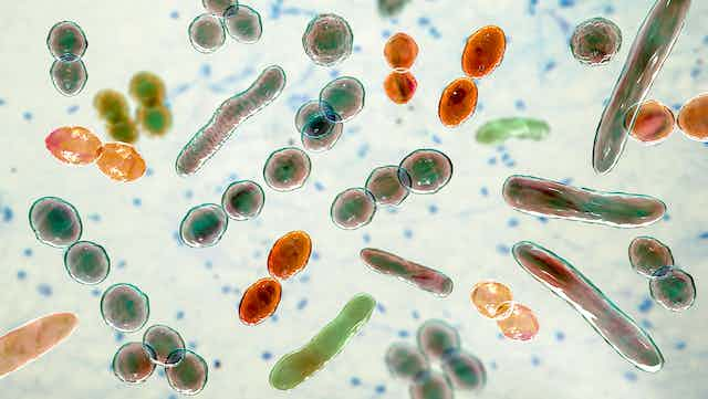
Cross-country culinary microbes: Uncovering a shared kitchen microbiota across European homes
2 June, 2023
University of Louisville researchers receive $5.8 million to prevent immune system dysregulation
2 June, 2023
The Future of Food Safety: Detecting Pathogens Before They Cause Illness
1 June, 2023
Why do some people live to be 100? Intestinal bacteria may hold the answer
Next Page
→
Subscribe
Subscribed
Microbiome & Microbiology Review
Join 111 other subscribers
Sign me up
Already have a WordPress.com account?
Log in now.
Microbiome & Microbiology Review
Edit Site
Subscribe
Subscribed
Sign up
Log in
Report this content
View site in Reader
Manage subscriptions
Collapse this bar