Tag Archives: Cell Death

Study provides evidence for a strong role of autophagy in controlling intracellular infections

Researchers at the Francis Crick Institute have found that the body’s process of removing old and damaged cell parts, is also an essential part of tackling infections that take hold within our cells, like TB.

If this natural process can be harnessed with new treatments, it could present an alternative to, or improve use of antibiotics, especially where bacteria have become resistant to existing drugs.

In their study, published in Nature Microbiology today, ahead of World TB Day on the 24th March, the team studied genes key to bacteria’s ability to evade autophagy, a pathway that cells use to destroy themselves when they are under stress or infected.

They engineered human immune cells called macrophages from specialist stem cells called induced pluripotent stem cells, which have the ability to become any cell type in the body. They then used genome editing tools to manipulate the macrophages ability to perform autophagy. When genes key to autophagy were removed and the cells were infected with Mycobacterium tuberculosis (bacilli that cause TB), the bacterial infection took hold, replicating more within the engineered cells and causing mass host cell death.

These results are evidence for a strong role of autophagy in controlling intracellular infections like TB. If this pathway can be boosted or strengthened, it could be a new avenue for tackling antibiotic resistance, by making existing antibiotic drugs more effective or presenting an alternative to drugs in cases where bacteria have evolved resistance.

I first studied the role of autophagy in infection during my PhD, so it’s incredible to see renewed interest in this field. Using the latest technologies, we’ve been able to show a key role for this pathway in controlling infection.

As immunotherapies have harnessed the immune system to fight cancer, boosting this immune defense with a host-directed therapy, could be a valuable new tool in the fight against infections, particularly those becoming resistant to antibiotics.”

Max Gutierrez, Head of the Host-Pathogen Interactions in Tuberculosis Laboratory at Francis Crick Institute

The team also validated their results using macrophages isolated from blood samples, confirming the importance of autophagy in human defenses.

Beren Aylan, joint first author and PhD student at the Crick together with Elliott Bernard and Enrica Pellegrino, said: “Antibiotic resistance is a huge threat to our health so it’s incredibly important to understand how our bodies fight infection and where there might be room for improvement.

“TB is a great example of where targeting our own immune defenses could be really effective, because it takes a very long course of different antibiotic treatments to effectively remove the infection. Anything that can be done to more effectively remove bacteria, could also make a huge difference to the cost and accessibility of treatments.”

The team are now planning to screen for drug compounds that could be used to boost autophagy in a targeted way.

“Boosting the autophagy pathway isn’t as simple as it might seem,” adds Max. This is because all parts of the body use autophagy as a way to recycle old and damaged cells. In order to safely increase autophagy in the location of infections, we need to target the pathway in macrophages alone.”

Source:
Journal reference:

Aylan, B., et al. (2023). ATG7 and ATG14 restrict cytosolic and phagosomal Mycobacterium tuberculosis replication in human macrophages. Nature Microbiology. doi.org/10.1038/s41564-023-01335-9

Simple blood tests for telomeric protein could provide a valuable screen for certain cancers

Once thought incapable of encoding proteins due to their simple monotonous repetitions of DNA, tiny telomeres at the tips of our chromosomes seem to hold a potent biological function that’s potentially relevant to our understanding of cancer and aging.

Reporting in the Proceedings of the National Academy of Science, UNC School of Medicine researchers Taghreed Al-Turki, PhD, and Jack Griffith, PhD, made the stunning discovery that telomeres contain genetic information to produce two small proteins, one of which they found is elevated in some human cancer cells, as well as cells from patients suffering from telomere-related defects.

Based on our research, we think simple blood tests for these proteins could provide a valuable screen for certain cancers and other human diseases. These tests also could provide a measure of ‘telomere health,’ because we know telomeres shorten with age.”

Jack Griffith, PhD, the Kenan Distinguished Professor of Microbiology and Immunology and Member of the UNC Lineberger Comprehensive Cancer Center

Telomeres contain a unique DNA sequence consisting of endless repeats of TTAGGG bases that somehow inhibit chromosomes from sticking to each other. Two decades ago, the Griffith laboratory showed that the end of a telomere’s DNA loops back on itself to form a tiny circle, thus hiding the end and blocking chromosome-to-chromosome fusions. When cells divide, telomeres shorten, eventually becoming so short that the cell can no longer divide properly, leading to cell death.

Scientist first identified telomeres about 80 years ago, and because of their monotonous sequence, the established dogma in the field held that telomeres could not encode for any proteins, let alone ones with potent biological function.

In 2011 a group in Florida working on an inherited form of ALS reported that the culprit was an RNA molecule containing a six-base repeat which by a novel mechanism could generate a series of toxic proteins consisting of two amino acids repeating one after the other. Al-Turki and Griffith note in their paper a striking similarity of this RNA to the RNA generated from human telomeres, and they hypothesized that the same novel mechanism might be in play.

They conducted experiments – as described in the PNAS paper – to show how telomeric DNA can instruct the cell to produce signaling proteins they termed VR (valine-arginine) and GL (glycine-leucine). Signaling proteins are essentially chemicals that trigger a chain reaction of other proteins inside cells that then lead to a biological function important for health or disease.

Al-Turki and Griffith then chemically synthesized VR and GL to examine their properties using powerful electron and confocal microscopes along with state-of-the-art biological methods, revealing that the VR protein is present in elevated amounts in some human cancer cells, as well as cells from patients suffering from diseases resulting from defective telomeres.

“We think it’s possible that as we age, the amount of VR and GL in our blood will steadily rise, potentially providing a new biomarker for biological age as contrasted to chronological age,” said Al-Turki, a postdoctoral researcher in the Griffith lab. “We think inflammation may also trigger the production of these proteins.”

Griffith noted, “When you go against current thinking, you are usually wrong because you are bucking many people who’ve worked so diligently in their fields. But occasionally scientists have failed to put observations from two very distant fields together and that’s what we did. Discovering that telomeres encode two novel signaling proteins will change our understanding of cancer, aging, and how cells communicate with other cells.

“Many questions remain to be answered, but our biggest priority now is developing a simple blood test for these proteins. This could inform us of our biological age and also provide warnings of issues, such as cancer or inflammation.”

Source:
Journal reference:

Al-Turki, T., et al. (2023) Mammalian Telomeric RNA (TERRA) can be translated to produce valine-arginine and glycine-leucine dipeptide repeat proteins. PNAS. doi.org/10.1073/pnas.2221529120.

Altered gut microbiome plays a major role in the progression of endometriosis in animal model

About 196 million women worldwide suffer from endometriosis, a condition that typically causes pelvic pain and infertility. Endometriosis develops when lining inside the womb grows attached to surrounding tissues, such as the intestine or the membrane lining the abdominal cavity, causing bleeding, pain and other symptoms. Despite decades of research, little is known about the factors that contribute to the development of endometriosis.

Evidence suggests that the microbiome, a community of microorganisms living inside the body, is altered in women with endometriosis. In this study published in the journal Cell Death & Discovery, researchers at Baylor College of Medicine discovered that an altered gut microbiome plays a pivotal role in endometriosis disease progression in an animal model.

“To investigate the role of the microbiome in endometriosis we first implemented a novel mouse model of the condition in which we eliminated the microbiome using antibiotics,” said lead author Dr. Rama Kommagani, associate professor in the Departments of Pathology and Immunology and of Molecular Virology and Microbiology at Baylor.

The researchers found that mice lacking gut microbiome had smaller endometriotic lesions than mice with a microbiome. Furthermore, when gut microbiome-free mice received gut microbiota from mice with endometriosis, the lesions grew as large as those in mice retaining their microbiome. These findings suggest that altered gut bacteria drive disease progression. On the other hand, the uterine microbiome did not seem to affect disease progression.

The team also discovered a novel signature of microbiome-derived metabolites, products produced by the microbes, that were significantly altered in feces of mice with endometriosis. Supporting the role of microbiome metabolites in disease progression, Kommagani and his colleagues found that treatment of endometriotic cells and mice with the metabolite called quinic acid significantly enhanced the cellular proliferation and endometriotic lesion growth, respectively.

The findings suggest that certain microbiome communities and/or their metabolites can contribute to endometriosis progression and that modifying the composition of these communities could help control the condition in human patients. “We are currently investigating this possibility,” Kommagani said.

The findings also suggested that studying microbiome metabolites in human stool samples could be used as a diagnostic tool. “Endometriosis is typically diagnosed with ultrasound, and an invasive procedure is necessary to characterize the lesion well,” Kommagani said. “We are investigating whether microbiome metabolites in human stool samples could be a useful diagnostic tool and also whether some of these metabolites could be used as a treatment strategy.”

Women with endometriosis also tend to have bowel issues, such as colitis or inflammatory bowel syndrome.

We are interested in determining whether changes in the gut microbiome could affect bowel conditions and the possibility of controlling them by modifying the microbiome or with their metabolites.”

Dr. Rama Kommagani, Lead Author

Source:
Journal reference:

Chadchan, S.B., et al. (2023) Gut microbiota and microbiota-derived metabolites promotes endometriosis. Cell Death Discovery. doi.org/10.1038/s41420-023-01309-0.

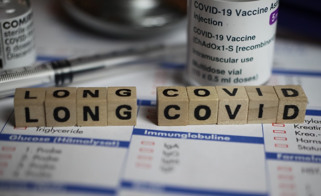
What are the major findings of long COVID research?

In a recent review published in Nature Reviews Microbiology, researchers explored existing literature on long coronavirus disease (COVID). They highlighted key immunological findings, similarities with other diseases, symptoms, associated pathophysiological mechanisms, and diagnostic and therapeutic options, including coronavirus disease 2019 (COVID-19) vaccinations.

Study: Long COVID: major findings, mechanisms and recommendations. Image Credit: Ralf Liebhold/Shutterstock

Long COVID refers to a multisystemic disease among SARS-CoV-2 (severe acute respiratory syndrome coronavirus 2)-positive individuals, with increasing prevalence rates by the day. Studies have reported on long COVID risk factors, symptoms, pathophysiology, diagnosis, and treatment options, with increasing similarities between long COVID and other diseases such as POTS (postural orthostatic tachycardia syndrome) and ME/CFS (myalgic encephalomyelitis/ chronic fatigue syndrome).

About the review

In the present review, researchers explored the existing data on long COVID immunology, symptoms, pathophysiology, diagnosis, and therapeutic options.

Key long COVID findings and similarities with other diseases

Studies have reported persistently reduced exhausted T lymphocytes, dendritic cells, cluster of differentiation 4+ (CD4+) lymphocyte and CD8+ lymphocyte counts, and greater PD1 (programmed cell death protein-1) expression. In addition, increase in innate cell immunological activities, non-classical monocytes, expression of interferons (IFNs)-β, λ1, and interleukins (IL)-1β, 4,6, tumor necrosis factor (TNF). Cytotoxic T lymphocyte expansion has been linked to gastrointestinal long COVID symptoms, and persistent increase in CCL11 (C-X-C motif chemokine 11) expression has been linked to cognitive dysfunction among long COVID patients.

Elevated autoantibody titers have been reported among long COVID patients, such as autoantibodies against ACE2 (angiotensin-converting enzyme 2), angiotensin II receptor type I (AT1) receptors, β2-adrenoceptors, angiotensin 1–7 Mas receptors, and muscarinic M2 receptors. Reactivation of Epstein-Barr virus (EBV) and human herpes virus-6 (HHV-6) has been reported in long COVID patients and ME/CFS. EBV reactivation has been linked to neurocognitive impairments and fatigue in long COVID.

SARS-CoV-2 persistence reportedly drives long COVID symptoms. SARS-CoV-2 proteins and/or ribonucleic acid (RNA) have been detected in cardiovascular, reproductive, cranial, ophthalmic, muscular, lymphoid, hepatic, and pulmonary tissues, and serum, breast, urine, and stool obtained from long COVID patients. Similar immunological patterns are noted between long COVID and ME/CFS, with elevated cytokine levels in the initial two to three years of disease, followed by reduction with time, without symptomatic improvements in ME/CFS. Lower cortisol levels, mitochondrial dysfunction, post-exertional malaise, dysautonomia, mast cell activation, platelet hyperactivation, hypermobility, endometriosis, menstrual alterations, and intestinal dysbiosis occur in both conditions.

Long COVID symptoms and underlying pathophysiological mechanisms

Long COVID-associated organ damage reportedly results from COVID-19-induced inflammation and associated immune responses. Cardiovascular long COVID symptoms such as chest pain and palpitations have been associated with endothelial dysfunction, micro-clotting, and lowered vascular density. Long COVID has been associated with an increased risk of renal damage and type 2 diabetes. Ophthalmic symptoms of long COVID, including altered pupillary responses to light, result from the loss of small nerve fibers in the cornea, increased dendritic cell density, and impaired retinal microvasculature. Respiratory symptoms such as persistent cough and breathlessness result from altered pulmonary perfusion, epithelial injury, and air entrapment in the airways.

Cognitive and neurological long COVID symptoms include loss of memory, cognitive decline, sleep difficulties, paresthesia, balancing difficulties, noise and light sensitivity, tinnitus, and taste and/or smell loss. Underlying pathophysiological mechanisms include kynurenine pathway activation, endothelial injury, coagulopathy, lower cortisol levels, loss of myelin, microglial reactivation, oxidative stress, hypoxia, and tetrahydrobiopterin deficiency.  Gastrointestinal symptoms such as pain in the abdomen, nausea, appetite loss, constipation, and heartburn have been associated with elevated Bacteroides vulgatus and Ruminococcus gnavus counts and lower Faecalibacterium prausnitzii counts. Neurological symptoms often have a delayed onset, worsen with time and persist longer than respiratory and gastrointestinal symptoms, and long COVID presents similarly in children and adults.

Diagnostic and therapeutic options for long COVID, including COVID-19 vaccines

The diagnosis and treatment of long COVID are largely symptom-based, including tilt tests for POTS, magnetic resonance imaging (MRI) to detect cardiovascular and pulmonary impairments, and electrocardiograms to detect QRS complex fragmentation. Salivary tests and serological tests, including red blood cell deformation, lipid profile, complete blood count, D-dimer, and C-reactive protein (CRP) evaluations, can be performed to assess immunological biomarker levels. PCR (polymerase chain reaction) analysis is used for SARS-CoV-2 RNA detection and quantification, and antibody testing is performed to assess humoral immune responses against SARS-CoV-2.

Pharmacological treatments include intravenous Ig for immune dysfunction, low-dosage naltrexone for neuronal inflammation, beta-blockers for POTS, anticoagulants for microclot formation, and stellate ganglion blockade for dysautonomia. Other options include antihistamines, paxlovid, sulodexide, and pycnogenol. Non-pharmacological options include cognitive pacing for cognitive impairments, diet limitations for gastrointestinal symptoms, and increasing salt consumption for POTS. COVID-19 vaccines have conferred minimal protection against long COVID, the development of which depends on the causative SARS-CoV-2 variant, and the number of vaccination doses received. Long COVID has been reported more commonly post-SARS-CoV-2 Omicron BA.2 subvariant infections.

Based on the review findings, long COVID is a multiorgan disease that has debilitated several lives worldwide, for which diagnostic and therapeutic options are inadequate. The findings underscored the need for future studies, clinical trials, improved education, mass communication campaigns, policies, and funding to reduce the future burden of long COVID.

Journal reference:

Our immune system has to be able to rapidly mount a response to pathogenic invaders, wounds, and other …

Our immune system has to be able to rapidly mount a response to pathogenic invaders, wounds, and other injuries. But it also has to be carefully controlled. When the body overreacts to an infection, it can cause a condition known as sepsis, which can be deadly. Sepsis is estimated to have caused about 11 million deaths around the world in 2017 alone. Now, scientists are learning more about the immune molecules and cells that trigger sepsis. The findings have been reported Science Immunology.

Image credit: Pixabay

Cytokines are immune signaling molecules. Tumor necrosis factor (TNF) is a widely studied cytokine that has many roles. TNF is activated by a crucial component of bacterial cell walls, called lipopolysaccharide (LPS), and recruits immune cells to fight infection. TNF can aid in the repair of tissue and cell survival, but it also has to be regulated. Overactive TNF signaling has been implicated in inflammatory diseases like rheumatoid arthritis. The uncontrolled production of cytokines, including TNF, can also cause a cytokine storm, a dangerous situation.

There is no standardized definition for a cytokine storm, in part because it can be difficult to define the line between a normal and abnormal immune response. But there are commonly accepted clinical features of cytokine storms, such as fever, headache, fatigue, anorexia, rash, diarrhea, and respiratory symptoms that often progress to more serious problems including, but not limited to catastrophic hemorrhages, shock, hypoxemia, renal failure, or liver damage.

While scientists have developed TNF blockers that have been useful for some autoimmune disorders including rheumatoid arthritis, those drugs don’t prevent cytokine storms that arise in conditions like severe COVID-19 cases or sepsis. The mechanisms of TNF are still not well-understood.

In the new study, researchers induced death in mice from TNF. But when mice did not express either of two proteins that are linked to the immune response to LPS, called TRIF and CD14, survival improved. Previous work has shown that TRIF and CD14 help regulate a protein complex that is related to LPS-induced inflammation and cell death.

Blood cells called myeloid cells are known to produce a lot of TNF. The research also showed that if mice lacked neutrophils and macrophages, two kinds of myeloid cells, symptoms of sepsis were reduced and survival improved when mice were given a lethal dose of TNF. The study authors suggested that macrophages and neutrophils are, therefore, contributing significantly to TNF-mediated death in mice.

It may also be possible to treat sepsis by targeting TRIF and CD14, which could reduce cell death and inflammation.

Sources: New England Journal of Medicine, Medical Xpress via The Conversation, Science Immunology


Carmen Leitch

Study identifies Δ42PD-1 as novel therapeutic target for hepatocellular carcinoma immunotherapy

HKUMed researchers at AIDS Institute, Department of Microbiology and Department of Surgery, School of Clinical Medicine, and School of Biomedical Sciences discover the role of an isoformic programmed cell death protein 1 (PD-1), namely Δ42PD-1, in suppressing the function of killer T cells, which is a type of immune cells essential for killing cancer cells among hepatocellular carcinoma (HCC) patients. The study is a breakthrough because it demonstrates that Δ42PD-1 causes stronger functional loss of killer T cells, revealing a molecular mechanism underlying the failure of PD-1-targeted immune checkpoint blockade (ICB) therapy. Moreover, antibody drug targeting at Δ42PD-1 inhibits HCC progression in animal models, which is independent of the PD-1 pathway. The full research article is now published online in the journal of Gut, a top-tier academic journal.

Background

It is well known that HCC accounts for up to 92.3% of liver cancer cases in China. The 2018 Nobel Prize in Physiology or Medicine was awarded for the discovery of cancer ICB therapy by inhibition of negative immune regulation using PD-1-targeted antibody, such as Nivolumab. The ICB therapy has resulted in prolonged survival and even cure in some cancer patients. The ICB therapy, however, is not effective for about 80% of HCC patients. Understanding the mechanism of unsuccessful ICB, therefore, would be essential for discovering a novel therapeutic target to save more lives of HCC patients.

Research methods and findings

The research team found that human T cells, which express Δ42PD-1 but not PD-1, account for up to 71% of killer T cells in untreated HCC patients. Δ42PD-1 positive T cells are mainly found in tumor tissues, associated significantly with HCC poor prognosis. Moreover, Δ42PD-1 positive T cells have weaker killing function than PD-1 positive T cells. Treatment of HCC patients using Nivolumab, the PD-1-targeted ICB drug, even increases the number of Δ42PD-1 positive T cells, especially in patients with tumor progression. We demonstrated that Δ42PD-1 positive T cells inside tumors promote HCC growth through activating toll-like receptors-4-mediated inflammation. Instead of Nivolumab, anti-Δ42PD-1 antibody inhibits tumor growth in three HCC/humanized murine models through blocking of the Δ42PD-1-TLR4 axis, reducing the number of Δ42PD-1 positive T cells and increasing functional killer T cells inside tumor. These findings not only revealed a mechanism underlying the unsuccessful PD-1-targeted ICB therapy but also identify Δ42PD-1 as a novel therapeutic target for HCC immunotherapy.

Significance of the study

This important discovery has provided scientific evidence that Δ42PD-1 may serve as a novel drug target against HCC or other relevant cancers and may warrant the clinical development of a humanized Δ42PD-1-specific antibody for immunotherapy against HCC and related human cancers/diseases.

‘We were the first research group discovering the Δ42PD-1 protein in the world’, commented by Professor Chen Zhiwei, Director of AIDS Institute and Professor of the Department of Microbiology, School of Clinical Medicine, HKUMed, who led the study. ‘In this study, we not only further discover the dual activities of Δ42PD-1 on human T cells in both suppressing anti-tumor immune response and promoting tumorigenesis but also generate a potential anti-Δ42PD-1 antibody drug for HCC treatment’.

‘Besides immunotherapy against HCC, the anti-Δ42PD-1 antibody can also be used as a drug to prevent HCC recurrence without induction of graft rejection after liver transplantation’, added by Professor Nancy Man Kwan, Department of Surgery, School of Clinical Medicine, HKUMed.

About the research team

The collaborative research team was led by Professor Chen Zhiwei, Director of AIDS Institute and Professor of the Department of Microbiology, School of Clinical Medicine, HKUMed, together with Professor Nancy Man Kwan, Department of Surgery, School of Clinical Medicine, HKUMed and Dr Tan Zhiwu, research assistant professor at AIDS Institute and Department of Microbiology, School of Clinical Medicine, HKUMed. This collaborative team includes Chiu Mei-sum, Dr Zhou Dongyan, Yan Chi-wing, Kwan Ka-yi, Dr Wong Yik-chun, Li Xin, Dr Li Liu from AIDS Institute and Department of Microbiology, School of Clinical Medicine, HKUMed; Dr Yang Xinxiang, Dr Cheung Tan-to, Dr Wang Yuewen, Dr Zhu Jiye, Professor Lo Chung-mau, Department of Surgery, School of Clinical Medicine, HKUMed; Dr Yue Ming and Dr Song Youqiang from School of Biomedical Sciences, HKUMed; and Dr Anthony Chan Wing-hung, Dr Zhou Jingying, Professor To Ka-fai, Professor Alfred Cheng Sze-lok, Professor Stephen Lam Chan from the Chinese University of Hong Kong.

Source:
Journal reference:

Tan, Z., et al. (2022) Isoformic PD-1-mediated immunosuppression underlies resistance to PD-1 blockade in hepatocellular carcinoma patients. Gut. doi.org/10.1136/gutjnl-2022-327133.