Microbiom News
News
Microbiology
Microbiome
Health u0026amp; Nutrition
Diseases u0026amp; Disorders
Infectious diseases
Product Review
MARINE MICROBIOLOGY
ANTIMICROBIALS
VIRAL INFECTION
EPIDEMIOLOGY
TOPICS
Microbiome
Health u0026amp; Nutrition
Diseases u0026amp; Disorders
Infectious diseases
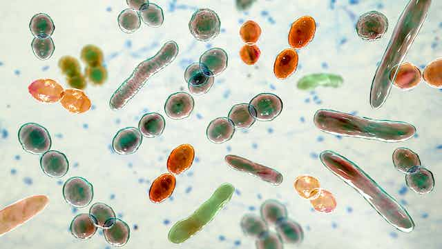
Bacteria
Virus
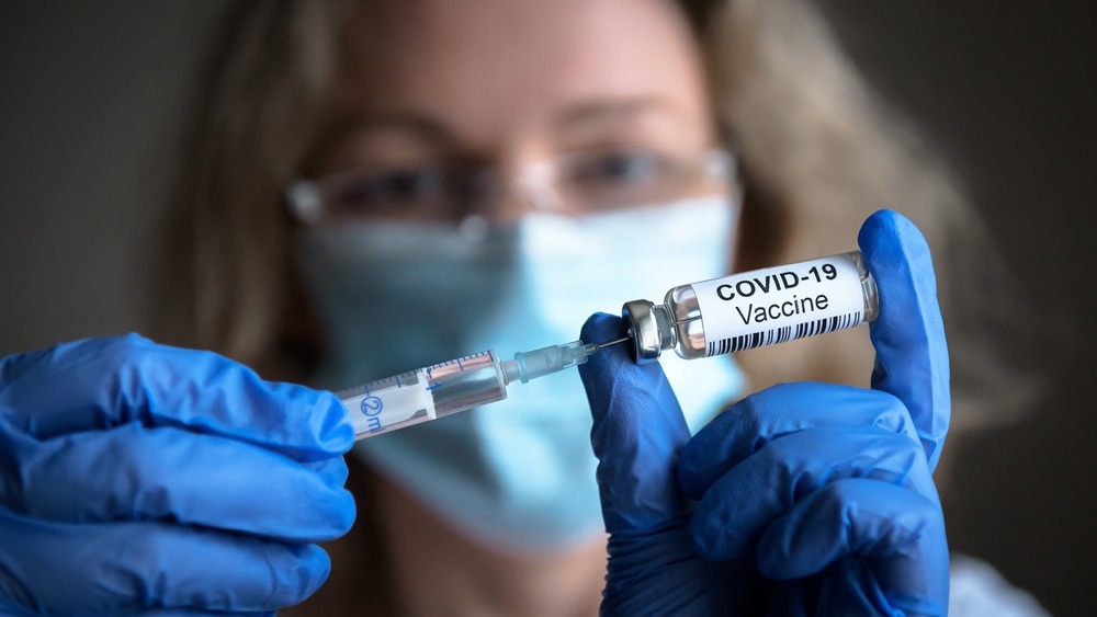
COVID-19
antibiotic resistance
Gastroenterology
Pathogen
Microbials
Antibiotics
Vaccinations
Microbiome
Health & Nutrition
Diseases & Disorders
Product Review
Newsletter
Contact
Facebook
Instagram
TikTok
Pinterest
YouTube
Twitter
Microbiome & Microbiology Review
Search
Drugs
9 June, 2023
Global study provides high-quality data to improve treatment of newborn babies with sepsis
5 June, 2023
UVA discoveries highlight the different roles of blood vessels in solid tumors
31 May, 2023
Experimental decoy provides long-term protection from SARS-Cov-2 infection
26 May, 2023
Antiviral drugs may be a new treatment strategy in the fight against Candida auris
22 May, 2023
Honokiol inhibits replication of SARS-CoV-2 in several cell types
19 May, 2023
Transforming antibiotic resistance testing: a novel, rapid and affordable technique
17 May, 2023
Long-ignored antibiotic could help fight against multi-drug resistant bacteria
15 May, 2023
New compound with antibacterial activity shows promising results within one hour in laboratory trials
13 May, 2023
Study identifies key genetic mechanism of drug resistance in the deadliest malaria parasites
8 May, 2023
Anticoronavirals: the development of COVID-19 therapies and the challenges that remain
Next Page
→
Subscribe
Subscribed
Microbiome & Microbiology Review
Join 111 other subscribers
Sign me up
Already have a WordPress.com account?
Log in now.
Microbiome & Microbiology Review
Edit Site
Subscribe
Subscribed
Sign up
Log in
Report this content
View site in Reader
Manage subscriptions
Collapse this bar