Microbiom News
News
Microbiology
Microbiome
Health u0026amp; Nutrition
Diseases u0026amp; Disorders
Infectious diseases
Product Review
MARINE MICROBIOLOGY
ANTIMICROBIALS
VIRAL INFECTION
EPIDEMIOLOGY
TOPICS
Microbiome
Health u0026amp; Nutrition
Diseases u0026amp; Disorders
Infectious diseases
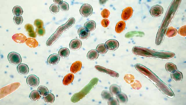
Bacteria
Virus
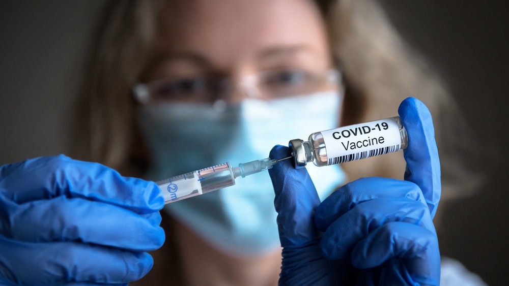
COVID-19
antibiotic resistance
Gastroenterology
Pathogen
Microbials
Antibiotics
Vaccinations
Microbiome
Health & Nutrition
Diseases & Disorders
Product Review
Newsletter
Contact
Facebook
Instagram
TikTok
Pinterest
YouTube
Twitter
Microbiome & Microbiology Review
Search
Molecule
10 June, 2023
UVA discovery advances efforts to prevent and better treat multiple sclerosis
8 June, 2023
UVA scientists discover a key determinant of multiple sclerosis risk
31 May, 2023
UNIGE researchers identify how the influenza A virus manages to penetrate host cells
31 May, 2023
Experimental decoy provides long-term protection from SARS-Cov-2 infection
26 May, 2023
Study highlights two strategies used by Salmonella to escape the human body’s defenses
17 May, 2023
Novel antibodies target human receptors to neutralize SARS-CoV-2 variants and future sarbecoviruses
15 May, 2023
New compound with antibacterial activity shows promising results within one hour in laboratory trials
8 May, 2023
Anticoronavirals: the development of COVID-19 therapies and the challenges that remain
24 April, 2023
Bioengineered drug candidate can counter S. aureus infection in early tests
4 April, 2023
Elucidating the function of BRCA2 gene offers insight into cancer development
Next Page
→
Subscribe
Subscribed
Microbiome & Microbiology Review
Join 111 other subscribers
Sign me up
Already have a WordPress.com account?
Log in now.
Microbiome & Microbiology Review
Edit Site
Subscribe
Subscribed
Sign up
Log in
Report this content
View site in Reader
Manage subscriptions
Collapse this bar