Microbiom News
News
Microbiology
Microbiome
Health u0026amp; Nutrition
Diseases u0026amp; Disorders
Infectious diseases
Product Review
MARINE MICROBIOLOGY
ANTIMICROBIALS
VIRAL INFECTION
EPIDEMIOLOGY
TOPICS
Microbiome
Health u0026amp; Nutrition
Diseases u0026amp; Disorders
Infectious diseases
Bacteria
Virus
COVID-19
antibiotic resistance
Gastroenterology
Pathogen
Microbials
Antibiotics
Vaccinations
Microbiome
Health & Nutrition
Diseases & Disorders
Product Review
Newsletter
Contact
Facebook
Instagram
TikTok
Pinterest
YouTube
Twitter
Microbiome & Microbiology Review
Search
Physiology
7 June, 2023
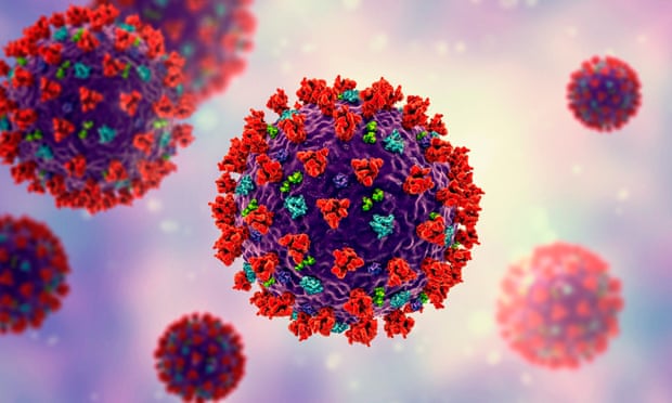
Two studies seek to understand the drivers of chronic and progressive phase of COVID-19
12 May, 2023
Scarring to the collagen framework causes dysfunction in Duchenne muscular dystrophy
10 May, 2023
Study may provide new avenues for addressing somatosensory symptoms of long COVID
19 April, 2023
Discovery offers a potential target for TB therapies
15 March, 2023
New Research Casts Fundamental Doubt on Long-Established Standard Model of Electroporation
10 March, 2023
New discoveries made regarding autism onset in mouse models
1 March, 2023
Gut-on-a-chip devices can bridge lab models and human biology
27 February, 2023
Rheumatoid arthritis (RA) is a complex, chronic inflammatory disease that is thought to affect about one percent of …
25 January, 2023
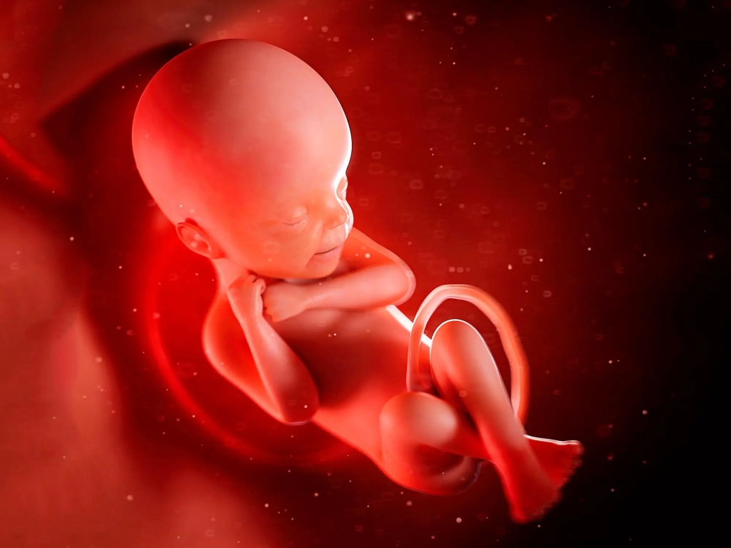
Experts Debunk Scientific Claims That Human Babies Are Colonized by Bacteria Before Birth
23 January, 2023
Gut feelings can be very real. There are neurons that connect the gut directly to the brain, and …
Next Page
→
Subscribe
Subscribed
Microbiome & Microbiology Review
Join 108 other subscribers
Sign me up
Already have a WordPress.com account?
Log in now.
Microbiome & Microbiology Review
Edit Site
Subscribe
Subscribed
Sign up
Log in
Report this content
View site in Reader
Manage subscriptions
Collapse this bar